
Digital Dental X-Rays in Warminster: Why They Matter for Early Detection and Treatment Planning
Published on Jul 22, 2026 | 15 minute read
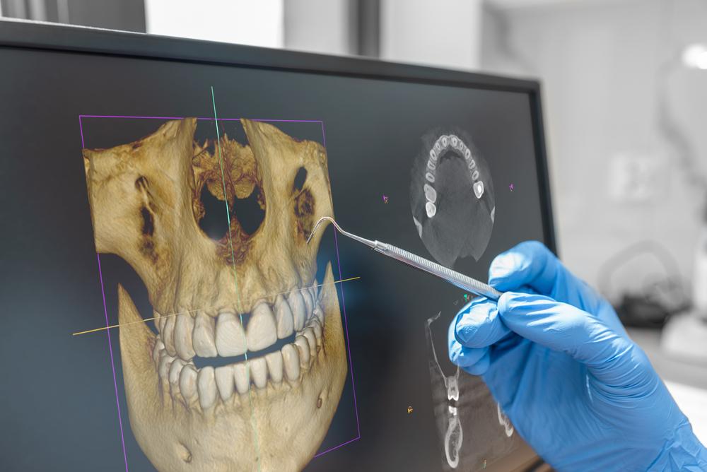
Have you ever wondered why a dentist recommends an X-ray when nothing in your mouth seems to hurt? The answer is simple: some dental problems begin in places that cannot be seen during a visual exam. Digital dental X-rays can reveal areas between teeth, below restorations, around tooth roots, and within the supporting bone, giving the dental team information that may not be visible from the surface.
For patients in Warminster, digital dental X-rays can make routine and problem-focused dental visits more informative. They help dentists look beyond what the eye can see, compare changes over time, and make treatment recommendations based on a more complete picture of the teeth and surrounding structures.
What Are Digital Dental X-Rays?
Digital dental X-rays are images created with electronic sensors rather than traditional photographic film. The sensor captures information that can be displayed on a computer screen shortly after the image is taken.
Because the image is digital, the dental team can enlarge specific areas, adjust the view, and compare current images with previous records. This can make it easier to discuss what is being seen and explain why a tooth may need monitoring or treatment.
Digital dental X-rays are only one part of diagnosis. They are used together with a clinical exam, your symptoms, dental history, and other findings to understand what is happening in the mouth.
Why a Visual Exam Cannot Show Everything
Dentists can learn a great deal by looking directly at teeth and gums, but some important areas are hidden.
For example, the contact point between two teeth is difficult to inspect directly. Decay may begin there before the outside of the tooth changes enough to be obvious. Likewise, the root of a tooth is covered by gum and bone, so problems around the root cannot always be seen without imaging.
Digital dental X-rays can help evaluate:
- Cavities between teeth
- Decay beneath or around some existing restorations
- Bone levels around teeth
- Tooth roots
- Developing or unerupted teeth
- Certain infections near the root
- Changes after dental treatment
- Areas involved in implant or restorative planning
Therefore, an X-ray can sometimes identify a concern before it creates a visible change or significant discomfort.
What Types of Dental X-Rays Might Be Recommended?
Not every dental visit requires the same images. The type of digital dental X-ray depends on what the dentist needs to evaluate.
Bitewing X-Rays
Bitewings commonly show the upper and lower back teeth in one area. They are useful for checking the spaces between teeth and monitoring the level of supporting bone.
These images can be especially helpful for finding cavities that are not visible during a normal exam.
Periapical X-Rays
A periapical image shows an entire tooth from the crown to the end of the root and includes surrounding bone. It may be recommended when a patient has pain, when the dentist is evaluating a particular tooth, or when the root and nearby structures need closer attention.
Panoramic Images
A panoramic image captures a broad view of the jaws, teeth, and surrounding structures. It may be useful for evaluating developing teeth, wisdom teeth, jaw relationships, or overall dental anatomy.
Three-Dimensional Imaging
Some treatment planning requires more detailed information than a standard two-dimensional image can provide. Three-dimensional dental imaging can be useful for selected implant, surgical, endodontic, and other complex cases because it shows anatomy from multiple angles.
The right image is chosen based on the clinical question. In other words, imaging should serve a purpose rather than being taken simply because a certain amount of time has passed.
Benefits of Digital Dental X-Rays
Digital dental X-rays offer several practical advantages as part of modern dental diagnosis and treatment planning. Depending on the situation, benefits may include:
- Earlier detection: Imaging can reveal certain cavities and changes before they become easy to see or feel.
- Better treatment planning: Dentists can evaluate roots, bone, tooth position, and hidden surfaces when planning restorative or surgical care.
- Easy comparison over time: Digital records make it possible to compare current images with earlier ones and monitor changes.
- Clear patient communication: Images can be displayed on a screen, enlarged, and discussed during the appointment.
- Efficient image capture: Digital systems can produce images quickly, which can help the dental team make decisions during the visit.
- Targeted monitoring: Specific areas can be followed when a dentist wants to watch a change rather than treat it immediately.
- Support for preventive care: When a problem is found earlier, treatment may be more conservative than it would be after the issue has progressed.
The main advantage is information. Digital dental X-rays help reduce guesswork by showing parts of the mouth that cannot be evaluated fully with vision alone.
Are Digital Dental X-Rays Safe?
Patients often ask about radiation exposure, and it is a reasonable question. Dental professionals are trained to use imaging carefully and to recommend X-rays based on individual need.
Modern digital systems are designed to capture diagnostic images efficiently. In addition, dental offices follow protective practices and use imaging only when the expected diagnostic value justifies it.
Your X-ray schedule may depend on several factors, including:
- Age
- Cavity history
- Current symptoms
- Gum health
- Previous dental treatment
- Presence of restorations
- Risk of future dental disease
- Whether you are a new or established patient
- The type of treatment being planned
Therefore, two patients may appropriately receive different imaging schedules.
If you are pregnant, may be pregnant, or have concerns about previous medical imaging, tell the dental team before X-rays are taken. That information helps the dentist make an informed decision about timing and necessity.
How Often Do You Need Dental X-Rays?
There is no universal schedule that fits every patient. Someone with a history of frequent cavities may need certain images more often than a person who has remained at low risk for years.
Likewise, a new patient may need imaging that helps establish a baseline and identify existing conditions. An established patient with stable oral health may need a different approach.
The decision is usually based on what the dentist needs to know. If there is no clinical reason for a particular image, it may not be necessary at that visit.
On the other hand, avoiding useful X-rays for too long can leave hidden problems undetected. The goal is to use imaging thoughtfully, not excessively and not inadequately.
Can X-Rays Find Cavities Before They Hurt?
Yes, certain cavities can be visible on dental X-rays before they create symptoms. This is especially important between back teeth, where early decay may be hidden from direct view.
Pain is not a reliable indicator of cavity size. A tooth can have progressing decay without hurting, particularly while the damage remains away from the nerve.
When decay is identified earlier, the dentist can determine whether the area needs treatment, preventive management, or monitoring. If a true cavity has formed and a restoration is needed, treating it earlier may help preserve more healthy tooth structure.
This is one reason digital dental X-rays can be valuable during preventive care even when you feel completely comfortable.
How Digital X-Rays Help With Tooth-Colored Fillings
When a cavity is suspected, digital dental X-rays can help show the location and approximate extent of the decay. That information can support decisions about whether a tooth-colored filling is appropriate.
For example, a small area between two teeth may not be visible from the chewing surface. A bitewing image can help the dentist see how far the decay appears to have progressed.
However, the final treatment decision is not made from the X-ray alone. The dentist also considers the clinical appearance of the tooth, the strength of the remaining structure, symptoms, and bite forces.
If the tooth has extensive damage or a large existing restoration, a crown may sometimes provide more appropriate protection than a filling.
How Digital X-Rays Help With Gum Health
X-rays do not show gum inflammation directly in the same way a clinical exam does. However, they can show the level of bone that supports the teeth.
This information is important when evaluating periodontal health. If bone levels have changed, the dentist can combine that information with gum measurements, bleeding patterns, tartar buildup, and other findings.
Therefore, dental imaging and periodontal examination work together. Neither replaces the other.
For patients with a history of gum disease, comparing images over time can be especially useful for monitoring stability.
How Digital Imaging Supports Dental Implant Planning
Dental implants depend on careful planning because the implant is placed within the jawbone. The dentist needs to understand the available bone and the location of nearby anatomy before treatment.
In selected cases, three-dimensional imaging can provide a detailed view of the area and help with implant positioning. This information can support planning for the final replacement tooth, not just the implant itself.
The process is important because implant dentistry involves both function and restoration. The final tooth needs to fit the bite, emerge naturally from the gumline, and work with the surrounding teeth.
Therefore, imaging is part of building a treatment plan that considers the full picture.
What Happens When an X-Ray Shows Something Unexpected?
Not every finding means immediate treatment is necessary. Sometimes an image reveals an area that simply needs to be monitored.
The dentist may compare the finding with older records, perform additional clinical tests, or recommend another type of image if more detail is needed.
For example, a questionable area around an old restoration might be watched if the tooth is stable and the finding has not changed. In another situation, symptoms and imaging together may indicate that treatment is appropriate.
This is why dental diagnosis is a process. The X-ray provides evidence, but the dentist interprets that evidence in the context of the entire patient.
Can You See Your Own Dental X-Rays?
One of the useful features of digital dentistry is that images can often be shown on a monitor during the visit. Ask your dentist to explain what you are seeing.
You may be able to identify:
- The bright outline of enamel
- Existing fillings or crowns
- Spaces between teeth
- Tooth roots
- Bone surrounding the teeth
- Areas the dentist wants to monitor
Understanding the image can make treatment recommendations feel less mysterious. Instead of simply hearing that a tooth has a problem, you can discuss where the concern is and how it affects the treatment plan.
Digital X-Rays and Emergency Dental Visits
When a tooth suddenly hurts, breaks, or becomes sensitive to pressure, a focused digital dental X-ray may help identify the source.
Pain can come from decay, a cracked tooth, inflammation around the root, a failing restoration, gum problems, or several other causes. Because different conditions can feel similar, an image may provide important clues.
However, some cracks and soft-tissue conditions do not show clearly on standard X-rays. Therefore, the dentist may also use bite tests, temperature tests, visual examination, and other methods to make a diagnosis.
The goal is to identify the cause rather than simply treat the symptom.
How to Prepare for Dental X-Rays
Most dental X-rays require little preparation. You may be asked to remove certain removable items near the head and neck, depending on the type of image being taken.
During a bitewing or periapical X-ray, a small sensor is positioned inside the mouth. You may be asked to bite gently to hold it in place while the image is captured.
If you have a strong gag reflex, jaw discomfort, or difficulty holding sensors, tell the dental team. There may be ways to adjust positioning and make the process easier.
Clear communication helps the appointment go more smoothly, especially if dental X-rays have been uncomfortable for you in the past.
See More Clearly With Modern Dental Imaging
Digital dental X-rays are not simply pictures taken during a dental visit. They are diagnostic tools that can reveal hidden areas, support early detection, improve treatment planning, and help your dentist monitor changes over time. When used thoughtfully, they add valuable information to the clinical exam and help create a more complete picture of your oral health.
If you have questions about digital dental X-rays or are due for a dental exam, Book an Appointment with Dolan Dental in Warminster, PA. Call (267) 961-5884 to schedule your visit and learn what imaging, if any, is appropriate for your current dental needs.
